CLOSE ×

Pathology is only as good as its laboratory. At Inotiv, we are committed to providing you with exceptional histology and histochemistry services in a timely manner. Our experienced staff are experts in routine and advanced laboratory techniques, including image analysis, and provide you with high production capabilities while ensuring quality is maintained throughout every process. We offer stand-alone and integrated customization and are dedicated to the success of your project.

Our histology service delivers the following:

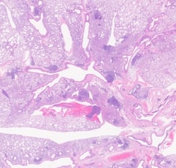
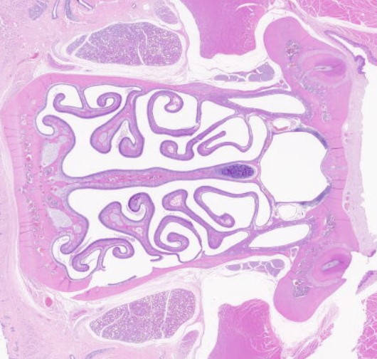
Histology Capabilities

Our specialized histology/pathology methods provide you with endpoint determinations that reflect tissue compartment localization and cell phenotype context. The outcome can then be further assessed through quantitative image analysis providing the data you need to make informed decisions. We are constantly developing new approaches to meet our clients' needs. If you do not see a capability you need listed below, please contact us.

  • GLP compliance
  • Robust QC and QA staffing
  • Routine tissue trimming, paraffin embedding, and sectioning of rodent and non-rodent tissues
  • Standard and oversized slides
  • H&E staining
  • Staining for brightfield and fluorescence microscopy
  • Wide selection of special stains
  • Serial and step sectioning
  • OCT embedding and cryosectioning for H&E, special, and Immunohistochemistry (IHC) staining
  • Customized bone decalcification methods
  • Extensive experience in:
    • Customized trimming and sectioning of bone, ocular, and OTICSpecific and complex brain tissue trimming and processing
    • Medical device support

HistoStain2-1

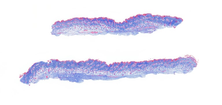
Stains and Applications

From routine applications to more complicated procedures, Inotiv has the expertise and the capability to determine the ideal specialty stain for your research needs. Our talented histotechnicians continuously create the highest quality novel stains that highlight what you need to move your program forward.


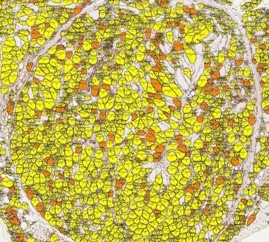
Image Analysis, TEM, and Quantitative Microscopy

At Inotiv, you’ll benefit from industry-recognized expertise in Image Analysis (IA). Consultation and design-based techniques, coupled with broad histochemical/immunohistochemical capabilities and knowledge of disease, ensure we deliver the best and most efficient quantitative evaluations possible to accelerate your development project.

We offer:

  • Computer assisted, high-throughput capability
  • Artificial Intelligence (AI) applied across multiple platforms, including Visiomorph™, Visiopharm®, inForm® and HALO®
  • Stereology
    • Unbiased, design-based, using statistically proven principles
    • Multiple platforms, including newCAST™
  • Conducted on brightfield whole slide scanned images (Aperio AT2) or IF imaged slides (Vectra® Polaris™)
  • HALO® and Visiopharm® image analysis platforms allow analysis of IHC and ISH with modular expansion to include H-scoring of membrane expressed tumor targets and spatial distancing
  • Multiplex IHC analysis
  • Cell proximity evaluation
  • Multiplex IF analysis conducted with inForm or Visiopharm platforms
  • Transmission electronic microscopy (TEM) is considered the gold standard for determining several important diagnoses such as:
    • Phospholipidosis 
    • Identification of immune deposits in the renal glomerulus
    • Endoplasmic reticulum stress
    • Neuroendocrine cell identification and more
  • Quantitative microscopy provides you with high-quality data for applications such as:
    • Muscle fiber cross-sectional area and Feret’s diameter
    • Co-expression of markers; double-stain positivity in the cytoplasm or nucleus
    • Area and area ratios; volume and volume ratios (e.g., volume of infarcted tissue, collagen, or mucus in an organ)
    • Membrane marker quantitation
    • Immune cells in tumors
    • Object quantitation (e.g., microvessels, amyloid plaque, glomeruli, islets)
    • Number of cells in a structure or organ
    • Microglial activation
    • Layer thickness
    • Proliferation assays
  • Reporting — pathologist oversight of prepared illustrative images and interpretive reports
Image-Analysis_yellow-edited

Whole Slide Scanning

Our team creates beautiful, extremely high-resolution whole slide images up to 40x magnification, which then can be seamlessly quantified using Visiopharm® or HALO® image analysis software to distribute data to you.

Our capabilities include:

  • Whole slide brightfield and fluorescence scanning of H&E, IHC, IF, ISH, and special stains
  • Diagnostic quality, high through-put resolution images up to 40x preserved digitally
  • Zoom in and out, pan around the scanned slide just like a microscope
  • Images are compatible with several third-party image-analysis providers
  • Whole slide images can be returned via hard drive, DVD, or reviewed online
  • Simple, secure online access from any web browser, on any computer
  • Evaluate scanned slides, organized by study, all in one place

WholeSlide-Skin-Control-Trichrome-scan_edited-768x363

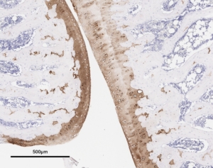
Immunohistochemistry (IHC) and Immunofluorescence (IF)

At Inotiv, we are committed to providing you with exceptional immunohistochemistry (IHC) and immunofluorescence lab services. Our extensive expertise with advanced techniques across a wide array of disease models allows us to produce IHC and IF staining that research and discovery organizations rely on for accurate assessments of their preclinical programs. Our ability to provide specialized approaches to your complex needs, while still maintaining quick turnaround times and high quality, makes Inotiv your partner of choice with the ideal source for reliable IHC and IF lab services.

Here’s how we can support you:

  • Method development
  • New antibody protocol optimization
  • Custom antibody panels
  • Immunophenotyping markers
  • Variety of cell type specific markers across multiple species
  • Transduced proteins and tags
  • Bone and other specialty tissues
  • Paraffin and frozen sections
  • Staining for Bright field and fluorescence microscopy
  • Antibody validation
  • Project specific R&D
  • Automated quantification of IHC including Nuclear, Membrane, Cytoplasm, and CD markers

Inotiv can help with other aspects of your project, such as method or protocol development and rushed completion requirements.

Collagen-II-Rat-Knee-B_IHC-300x237

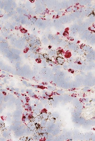
In Situ Hybridization (ISH)

We offer the ISH technique to bring you faster and more accurate results when quantifying your nucleic acid targets across tissues and cell lines.

This includes:

  • Custom-made probes to identify specific gene expression and miRNA, lncRNA, and viral DNA genomes
  • Frequently used as an adjunct to assess IHC validity
  • Multiplex staining
    • ISH combined with IHC capabilities
    • Multiplex FISH


MultiPlex-ISH-1

Optimized Antibody List

Below you can access our IHC antibody list. We can accommodate other specific or modified stains and/or procedures not listed. We are adding new antibodies weekly, so please contact us to request an antibody not listed.

Peer Review and Various Pathology Services

  • Evaluation of microscopic slides; our pathologists are available to travel or slides may be shipped to us
  • Full narrative pathology reports
  • Pathology data, to include organ and body weight correlations
  • Clinical pathology correlations to histopathology and interpretations
  • Pathology Working Groups
  • Consulting

If you do not see a capability you need, please contact us as we are regularly developing new models and processes to meet our clients' needs.

Subscribe to updates from Inotiv

Sign up and you’ll receive the latest updates that matter to you, delivered straight to your inbox.

Subscribe now